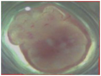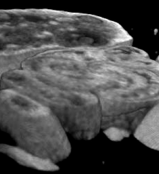
3D ex vivo imaging  - Whole tissue imaging - mouse ovary

Whole tissue imaging

Introduction

Novel Optical Coherent Technology (OCT) developed by SCREEN holdings has been used for morphological evaluation of complex 3D structures and tissues. The system is equipped with an 850-nm light from super luminescent diode (SLD). The OCT observation system adopts an inverted microscope, which picks up the reflected light from sample. Thereby enabling non-invasive detection of internal cavities and gaps in tissues.

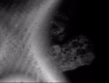
3D ex vivo imaging  - Whole tissue imaging - mouse ovary
Mouse ovary 3D cut view
3D ex vivo imaging  - Whole tissue imaging - mouse ovary - Given its tremendous use in clinical settings, SCREEN has adapted this technique in 3D ex vivo applications for detailed imaging of spheroids/ organoids and large tissues.
Ovary Cross section view 4 mm width
3D ex vivo imaging  - Whole tissue imaging - mouse ovary - Given its tremendous use in clinical settings, SCREEN has adapted this technique in 3D ex vivo applications for detailed imaging of spheroids/ organoids and large tissues.
Ovary Microscope 4 mm width

Presentation

Optical Coherent tomography (OCT) has been widely used in ophthalmology. It is a non-invasive imaging technology that renders the high resolution and cross-sectional images from retina.  Given its tremendous use in clinical settings, SCREEN has adapted this technique in 3D ex vivo applications for detailed imaging of spheroids/ organoids and large tissues. This technology allows to perform large tissue imaging, non-invasive monitoring of macro and sprouted neo-vasculature without the need for fluorescent staining for providing quantitative information about the vascular morphological changes. Our collaborator, Dr.Nobuo Nagai, Nagahama Institute of Biosciences and technology, and Dr. Tetsuya Ohbayashi from Totori University have successfully utilized Cell 3imager ESTIER for imaging of various tissues including Mouse ovary and kidney tissues.


  • 3D ex vivo imaging  - Whole tissue imaging - mouse ovary - Given its tremendous use in clinical settings, SCREEN has adapted this technique in 3D ex vivo applications for detailed imaging of spheroids/ organoids and large tissues. kidney 3D trimming view
    kidney 3D trimming view
  • 3D ex vivo imaging  - Whole tissue imaging - mouse ovary - Given its tremendous use in clinical settings, SCREEN has adapted this technique in 3D ex vivo applications for detailed imaging of spheroids/ organoids and large tissues. kidney 3D trimming view
    kidney 3D view

Related Products

Would you like additional information?

I agree to the terms and conditions

I agree to the GDPR

Need help finding the right product?
Get in touch
+1 (844) 454-3399