Cell3iMager Neo -  Tumor spheroids - High speed and bright-field 3D spheroid imaging system. Cell 3imager NEO is an inexpensive and high performance and fastest imaging system. In less than 2 minutes per plate, Neo can rapidly image and analyse 3D structures, such as mono / multicellular spheroids non-invasively.  The companion 3D optimized analysis software allows fast image capture and data analysis including diameter, area, volume, and circularity of individual spheroids and colony measurements. Compatible with all SBS cell culture plates, microfluidics chips, organ-on-chips formats.

High speed and bright-field 3D spheroid imaging system

View Video

Cell3iMager’s sister unit, Cell3iMager neo has a compact design enabling the reduction of footprint by 40%. In addition to the existing functions of Cell3iMager, this highly versatile 3D spheroid counter provides excellent performance and scalability with even faster scanning speed for a single well plate and its numerous new software.

Imaging spheroids using conventional or even automated microscopes is a slow, low throughput process. High content imaging systems overcome some of these throughput issues, but are comparatively expensive, and still suffer from slow image capture/processing, and image analysis software not optimized for 3D spheroids. The Cell3iMager neo overcomes these limitations, featuring high-resolution scanning (up to 9600 dpi) without compromising scan time. This minimizes ‘out of incubator’ time to avoid jeopardizing cell health during analysis.

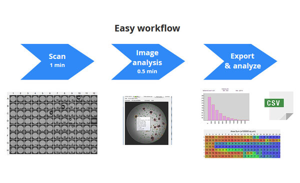
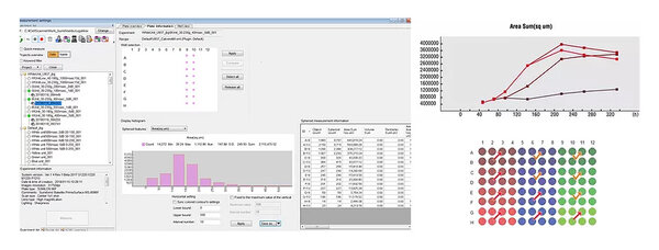
The powerful automated measurement and analysis software eliminates cumbersome manual morphological measurements and improves data reproducibility. Simply scan, measure, and analyze. Typical spheroid analysis includes a < 1 minute plate scan followed by a 20 to 30 second auto-measurement step. Data can be reviewed and processed in the software module, or exported for analysis in your preferred statistical software package. Rapid scan time (96-well plate in <1 minute) increases throughput (30 × 384-well plates/hour in the cc-5000 vs 2.5 plates/hour with conventional systems) Preset plate definitions for all major hanging drop, ULA, and micropatterned 3D cell culture platforms (6-, 12-, 24-, 48-, 96-, 384-well), dishes.

Key Applications

  • Phenotypic drug discovery
  • Drug sensitivity testing
  • Co-culture: loss of spheroid Area, volume or circularity
  • Combinatorial drug testing
  • Drug-target discovery and validation
  • Quality control
  • Toxicological evaluation using hepatocyte derived 3D spehroids
  • Label-free detection enables faster processing with fewer processes
  • Although adaptable to higher throughput plate readers, biochemical assays that monitor viability (e.g., ATP content) can effectively correlate to spheroid size, but are often lytic or otherwise destructive in nature, requiring multiple replicates to be run for longitudinal studies
  • Label-free measurement of changes in 3D microtissue size and morphology using the Cell3iMager enables efficient assessment of phenotypic endpoints without disrupting microtissue growth and can be used to focus selection of therapeutic targets and treatment strategies before costly and tedious testing in animal models
  • Drug sensitivity of HCT116 spheroids to the cytostatic drug Gemcitabine was tested in a time-course study, with spheroid size assessed daily for 1 week using the Cell3iMager
  • Gemcitabine inhibited growth at concentrations of 4 nM and higher, as displayed by plotting spheroid area in μm2 over time (left)
  • Comparison of day 7 spheroid area (black line, right) to total cellular ATP levels (green bars) at day 7 following treatment indicates spheroid size correlates closely with this lytic biochemical assay, while reducing spheroids used and processing time
  • Non-disruptive, continuous assessment of spheroid growth conserves precious cellular material, reduces reagent use, and saves handling time
Research field
  • 3D culture based-drug discovery and regenerative medicine research
  • Non-invasive research that researchers cannot fix/stain cultured cells
in vitro Drug-screening
High throughput
  • 54 second scan time per 1 micro well plate
  • 90 seconds for spheroids counting and quantifying area, diameter and pseudo volume for the spheroids derived from tumor cells or hepatocytes
Bright-field imaging
Entry model for 3D culture analysis
  • Extremely fast imaging system amenable to high throughput drug screening applications
  • Optimized for 3D culture analysis and bright-field imaging

1. High-throughput bright field scanners for 3D spheroids

kljölk

  • Cell3iMagers are bright field scanners designed specifically to simplify and accelerate quantitative and qualitative measurement of 3D spheroids.
  • This unique LED-based imaging system allows the user to identify and measure single or multiple spheroids per well in a microplate rapidly and automatically.
  • The companion 3D optimized analysis software allows faster image capture, measurement and data analysis

2. Z-stacking of 3D cellular structures

  • Cell3iMager has a focus composite function.
  • Merges stacking images of each Z-direction, for example gel culture or soft agar medium.
  • Measure/quantify mass of cells spread in the Z-direction at once

3. Compatible with various 3D culture microplates

  • Compatible with a wide variety of 3D culture microplates

4. Intelligent 3D software

  • Supports basic (single-time point) and time-course dose-response analysis, as well as a multi-planar (focus bracketing) scanning option to use with hydrogel-embedded spheroids
  • 30+ adjustable parameters for optimizing object identification, live/dead discrimination (e.g., bubbles, fibers, debris), and compensate for edge of well shadows and distortion as well as reduction of background noise
  • Identify and measure microtissues as in single spheroid per well or clonogenic assay formats (multiple spheroid colonies per well)
Well plates
Flat and U-shape bottom type well plate (6 to 384 well) and 35mm/ 60mm/100mm dish, microscope slide, T25-flask(only for CC-3000)
Plate number
CC-5000: 4 plates / CC-3000: 1 plate
Scan speed
CC-5000:54 seconds / 1 plate (2400dpi high speed mode) / CC-3000: 50 seconds / 1 plate (2400dpi high speed mode)
Automated masuring time
20 seconds / 1 plate (2400dpi high speed mode)
Scan resolution
200dpi, 300dpi, 600dpi, 1200dpi, 2400dpi, 4800dpi, 9600dpi
Light source / optical system
White LED / transmission illumination
Measuring
Number, area, pseudo volume, contour, optical density, circularity and edge sharpness
Output format
Captured image display and numerical data confirmation using software Granular distribution and proliferation trend chart confirmation Output of measured data in CSV file
Environment
Recommended temperture : 18 to 28℃ (64 to 82°F)
Power requirements
CC-5000:1Φ100 - 240V 0.12kw 0.9A / CC-3000: 1Φ100 - 240V 0.115kw 0.85A
Dimensions
CC-5000: W492 x D755 x H 294 mm / CC-3000: W594 x D 381 x H 294 mm
Weight
CC-5000: Approx. 40kg / CC-3000: Approx. 25kg
Cell3iMager Neo
 High speed and bright-field 3D spheroid imaging system

Would you like additional information?

I agree to the terms and conditions

I agree to the GDPR

Need help finding the right product?
Get in touch
+1 (844) 454-3399