
Size and morphology are important determinants to evaluate biological behavior of 3D microtissues, such as 3D multicellular tumor spheroids (MCTS). The SCREEN Cell3iMager, a bright-field spheroid scanner, provides a precise and fast analytical tool to perform growth and morphological profiling of microtissues. SCREEN has demonstrated a new application for the Cell3iMager in collaboration with Dr. Geoffrey Bartholomeusz and colleagues at MD Anderson Cancer Center.
Dr. Bartholomeusz’s group is focusing on developing 3D MCTS that could be used in high throughput RNAi and small molecule screens to identify novel targets for cancer therapy, to understand signaling pathways mediating the onset, tumor growth and invasion, as well as the mechanisms of action of effective anticancer agents.
His group has developed many relevant 3D systems to address important questions in respect to tumorigenesis and cancer treatment. As they continue to develop these complex 3D MCTS models, it becomes imperative that they have appropriate equipment and technology that enables determination of important end points for each study.
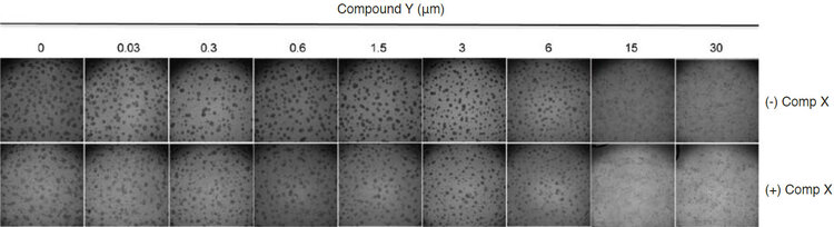
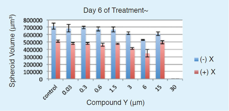
The Cell3iMager’s high quality optical system together with the easy-to-use preset assay parameters for quick data analysis and simple and intuitive software user interface affords the opportunity to scan 3D cultures with a high degree of resolution, and determine parameters such as area, diameter, volume, optical density and viability without interfering with the growth conditions of the cell culture. Plates can be simply removed from the incubator for a minimal amount of time to be scanned each day, enabling changes to 3D MCTS cultures in real time. An example of the versatility of this instrument is described below. In an ongoing study Dr. Bartholomeusz’s group is working towards understanding the mechanism of action of two natural compounds that have been demonstrated with relevant anti-tumor activities in clinical trials. In this study, spheroids were divided into two groups for a combinatorial treatment, in which group 1 was treated with different concentrations of Compound Y but not with Compound X. In Group 2, the MCTS were treated with Compound X (0.3 μm) as well as Compound Y at the same concentration range as of Ggroup 1. Both groups were scanned in the Cell3iMager from day 1 to day 6 post treatments and the average volume of the spheroids in each condition was determined. The data presented in figure below are the bright-field images (top) and quantitation of average spheroid volume (μm3) (bottom) on the final day of treatment (day 6).
This data suggests that the combination of the two compounds has a stronger effect on inhibiting growth of the 3D MCTS compared to treatment with single compound alone. The data generated from the Cell3iMager over the 6 day treatment period clearly indicated that compound X altered the morphology of the spheroids (data not shown) and sensitized the spheroids to Compound Y. This study also confirmed that the Compound X: Compound Y ratio (0.3 μm: 3.0 μm) was optimal in vitro, reflecting the ratio previously determined as optimal in the treatment of patients.
This data suggests that the combination of the two compounds has a stronger effect on inhibiting growth of the 3D MCTS compared to treatment with single compound alone. The data generated from the Cell3iMager over the 6 day treatment period clearly indicated that compound X altered the morphology of the spheroids (data not shown) and sensitized the spheroids to Compound Y. This study also confirmed that the Compound X: Compound Y ratio (0.3 μm: 3.0 μm) was optimal in vitro, reflecting the ratio previously determined as optimal in the treatment of patients.

, angiogenic vessels (neovasculature, sprout formation), gut model, skin models conveniently, by quantitating and monitoring the morphological changes occurring during the culture.")


