
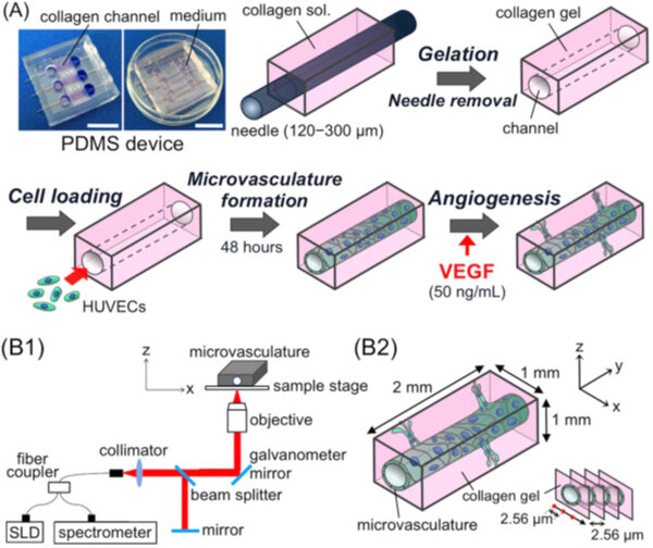
Takahashi et. al., 2017, have developed Three-dimensional (3D) in vitro microvasculature in a polydimethylsiloxane-based microdevice as a physiologically relevant model of angiogenesis. The angiogenic process is monitored using stage-top optical coherence tomography (OCT). OCT allows non-invasive monitoring of the 3D structures of the prepared host microvasculature and sprouted neovasculature without fluorescence staining. OCT monitoring takes only a few minutes to scan through the several-millimetre scale range, which provides the advantage of rapid observation of living samples. The obtained OCT crosssectional images capture 3D features of the angiogenic sprouting process and provide information on the dynamics of luminal formation. The stage-top system used in this study enables the observer to visualize the in vitro dynamics of 3D cultured cells simply and conveniently, offering an alternative monitoring method for studies on angiogenesis and providing quantitative information about vascular morphological changes.
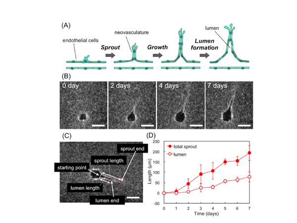

, angiogenic vessels (neovasculature, sprout formation), gut model, skin models conveniently, by quantitating and monitoring the morphological changes occurring during the culture.")


